
Suspicious Mole? Private vs NHS: What Happens and How Fast
A new or changing mole needs prompt assessment by someone trained to examine it with dermoscopy. On the NHS that means an urgent suspected cancer referral from your GP; privately at Midland Skin you book directly and are seen by a consultant dermatologist, with a biopsy arranged promptly if one is needed. This article explains what happens at each step on both routes and how long things usually take.
Key points
- Dermoscopy is the key first-line examination for a changing mole
- NHS – urgent GP referral, often triaged first by teledermatology
- Private – no referral needed, seen directly by a consultant dermatologist
- Biopsy technique is chosen to answer the question and limit scarring
- Suspicious moles are often removed at the same visit
- Results usually take about one to two weeks
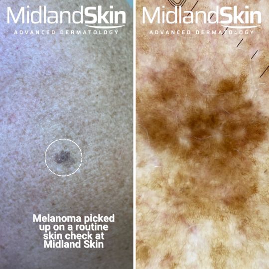
Melanoma pictures



The worry behind a changing mole and why speed matters
A new or changing mole can trigger understandable what-if thoughts. In clinic, we describe a lesion as suspicious when its pattern, colour, border or evolution suggests risk. The first-line tool is dermoscopy, a magnified and polarised light examination that improves accuracy over unaided visual inspection for identifying melanoma and ruling out false alarms.
Two routes, one goal: catching cancer early
NHS standards in plain English
If your GP is concerned about a mole, you are referred on the urgent suspected skin cancer pathway (previously called the “two-week wait”). In England this is measured against the Faster Diagnosis Standard, which aims to diagnose or rule out cancer within 28 days of referral, alongside 31-day and 62-day treatment standards. In many areas the first step is teledermatology: photographs, usually including dermoscopic images, are taken by a medical photographer or trained staff and reviewed remotely by a dermatologist. Depending on the images you may be reassured and discharged, booked straight in for a biopsy, or given a face-to-face clinic appointment where the lesion is examined with dermoscopy.
The private pathway at Midland Skin
Privately, you do not need a GP referral. You choose an appointment time that is prompt and suits you, and you are seen in person by a consultant dermatologist for a consultant-led dermoscopic assessment. If a biopsy is indicated, we discuss the most suitable technique, such as excision, punch or shave, and arrange it promptly under local anaesthetic with clear aftercare and a plan for results. Many suspicious moles are removed on the same day as the consultation where the consultant considers this appropriate; otherwise the procedure is booked promptly.
At a glance: NHS vs private for a suspicious mole
NHS route (typical)
- GP appointment and urgent suspected cancer referral
- Teledermatology: photographs and dermoscopic images taken and reviewed remotely by a dermatologist
- Hospital dermatology clinic if a face-to-face examination is needed
- Dermoscopy and decision: reassure, monitor or biopsy
- Biopsy on the day or booked
- Histology result and management plan
Measured against the 28-day Faster Diagnosis Standard.
Private route at Midland Skin (typical)
- Self-referral: book directly, no GP letter needed
- Consultant-led dermoscopic assessment in person
- Biopsy or excision under local anaesthetic, often at the same visit
- Histology result: most labs report within about one to two weeks, but timing varies by provider and laboratory workload
- Management plan
Your first assessment: what actually happens
We begin with your story: what changed, when you noticed it, any symptoms such as itch or bleeding, personal or family skin cancer history, and sun exposure. Then we examine the lesion and, when relevant, the surrounding skin. Dermoscopy reveals subsurface colours and structures that guide whether to watch, image or biopsy. Dermoscopy outperforms visual inspection alone.
Other moles can also be checked at your initial private consultation, and you can even opt to have a full head to toe skin check. You can find out more about our consultant-led mole checks here.
Teledermatology vs face-to-face
Good photographs, sometimes with dermoscopic images, can help triage and prioritise. Accuracy depends on image quality and clinical context. When there is uncertainty, an in-person dermoscopic assessment remains the safer choice.
Biopsy choices, scarring and safety: practical questions
- Excision removes the whole lesion with a margin. This is preferred when melanoma is suspected.
- Punch biopsy takes a core sample and is useful in specific diagnostic scenarios.
- Shave biopsy removes a superficial sample and is used selectively when appropriate.
All are done under local anaesthetic with clear wound-care advice. Any cut leaves a scar, yet careful planning of technique and closure helps minimise impact.
For a plain-English overview of what minor procedures involve under local anaesthetic, see Minor skin surgery. If the mole appears benign but causes problems such as snagging, catching or bleeding, we can discuss removal for comfort or function: Mole removal options.
Results and next steps: how long and what they mean
Most histopathology reports return in about two weeks. Timing varies with laboratory workload and whether any extra tests are needed. Your report guides the next step: reassurance and discharge if benign, or wider excision, further tests or ongoing monitoring if malignant or atypical.
Choosing NHS or private: a simple decision aid
- Risk and urgency – if melanoma is a concern, prioritise a prompt, high-quality dermoscopic assessment and a clear next step.
- Control and timing – if choosing a specific consultant dermatologist and a predictable appointment lowers anxiety and speeds decisions, private care may suit you.
- Complexity – previous melanoma, high-risk sites or multiple changing lesions often benefit from consultant-led review.
- Practicalities – consider travel, recovery time and help with aftercare.
- Continuity – you can move between routes; what matters is fast, expert assessment and clear communication.
Private mole checks: practical FAQs
Do I need a GP referral to book a private mole check?
No. You can book directly with Midland Skin without a referral.
Can the mole be removed on the same day as my consultation?
Often, yes. If your consultant recommends removing a suspicious mole, this is frequently done at the same visit under local anaesthetic. It depends on the assessment, so it cannot be promised in advance; where it is not appropriate on the day, the procedure is booked promptly. This is separate from our pre-arranged same-day procedure packages, which are for benign lesions such as skin tags and seborrhoeic keratoses.
Will the NHS remove a benign mole?
Generally, cosmetic benign lesions are not removed on the NHS. Suspicious lesions are assessed and biopsied if needed on a cancer pathway.
Can I switch between NHS and private?
Yes. You can have an assessment privately and still choose NHS or private treatment, providing the concern you seek treatment for is routinely provided in the NHS.
What should I bring to my appointment?
Any photographs you have of the mole over time, a note of when you first noticed the change, a list of your current medications, and details of any previous skin cancer or skin surgery. Please come without fake tan or make-up over the area.
This article provides general information only and is not a substitute for personalised medical advice. If you notice a fast-changing, bleeding or very dark lesion, arrange an urgent medical assessment.
References used for this article
- Dinnes J, Deeks JJ, Chuchu N, et al. Dermoscopy, with and without visual inspection, for diagnosing melanoma in adults. Cochrane Database Syst Rev 2018;12:CD011902. doi:10.1002/14651858.CD011902.pub2.
- Chuchu N, Dinnes J, Takwoingi Y, et al. Teledermatology for diagnosing skin cancer in adults. Cochrane Database Syst Rev 2018;12:CD013193. doi:10.1002/14651858.CD013193.
- NHS England. A teledermatology roadmap: implementing safe and effective teledermatology triage pathways and processes. 2023. england.nhs.uk
- NHS England. Faster diagnosis: the Faster Diagnosis Standard. england.nhs.uk
- National Institute for Health and Care Excellence. Melanoma: assessment and management (NG14). 2015, updated 2022. nice.org.uk
- Nasr I, McGrath EJ, Harwood CA, et al. British Association of Dermatologists guidelines for the management of adults with basal cell carcinoma 2021. Br J Dermatol 2021;185(5):899–920. doi:10.1111/bjd.20524.
- Keohane SG, Botting J, Budny PG, et al. British Association of Dermatologists guidelines for the management of people with cutaneous squamous cell carcinoma 2020. Br J Dermatol 2021;184(3):401–414. doi:10.1111/bjd.19621.
About the Author
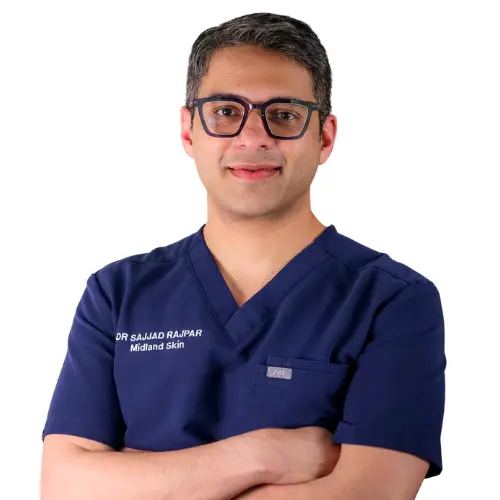
This page has been written and/or medically reviewed by Dr Sajjad Rajpar, Consultant Dermatologist and Medical Director of Midland Skin, Birmingham.
Dr Rajpar is on the GMC Specialist Register in Dermatology and has over 20 years’ experience in clinical dermatology. He provides clinical oversight to ensure the information on this page is accurate, balanced and consistent with current dermatology practice.
Dr Sajjad Rajpar
Date last updated: 26th Sep 2026
Book in with us
Expert, consultant led care in dermatology, skin surgery, eyelid surgery, lasers and aesthetics.

