
Basal Cell Carcinoma
Specialist assessment, diagnosis and treatment for basal cell carcinoma
At Midland Skin we offer Basal Cell Carcinoma consultations with a Dermatologist for patients in Birmingham, Solihull, Coventry, Wolverhampton, Worcester and the Midlands.


Quick Facts about basal cell carcinoma

Best for
Suspected or confirmed BCC

Appointment
Initial dermatology assessment

Treatment
Excision, Mohs or cream

Symptoms
Non-healing or bleeding lesion

Provider
Consultant dermatologist

Price
See below
What is basal cell carcinoma?
Basal cell carcinoma, usually shortened to BCC, is the most common form of skin cancer. It is sometimes also called a rodent ulcer.
BCC develops within the outer layer of the skin and is most often associated with ultraviolet exposure from sunlight or sunbeds. It commonly develops on sun-exposed areas such as the face, scalp, ears and neck, but it can occur anywhere on the body.
Most BCCs grow slowly and very rarely spread to other parts of the body. However, they continue to grow if left untreated and can extend more deeply into the surrounding skin and nearby structures. A larger or more deeply established BCC may require more extensive surgery and leave a larger scar.
Early assessment generally allows the BCC to be treated when it is smaller and easier to remove.
Who is more likely to develop BCC?
People are more likely to develop BCC if they:
- have fair skin that burns easily
- have had significant sun exposure through work, outdoor activities, holidays or sunbeds
- have previously had a BCC or another skin cancer
- are older, although BCC is increasingly diagnosed in younger adults
- have a weakened immune system
- have previously received radiotherapy or certain forms of medical ultraviolet treatment
- have one of the rare inherited conditions associated with multiple BCCs
People of every skin colour can develop skin cancer. A lesion should not be dismissed solely because someone does not have fair skin or the usual risk factors.
What does basal cell carcinoma look like?
BCC does not have one single appearance. It may look like a harmless spot, patch, scar or area of irritated skin.
Signs that can occur include:
- a scab that repeatedly bleeds, crusts and appears to heal before returning
- a pink, red, pearly or skin-coloured lump that gradually becomes larger
- a shiny bump with fine red blood vessels visible across its surface
- a flat, scaly pink or red patch that does not clear
- an ulcer or crater within the centre of a skin lesion
- a pale, firm or scar-like area with poorly defined edges
- a brown or black lesion that may resemble a mole
Nodular BCC
A nodular BCC often appears as a pink, pearly or translucent lump. Small blood vessels may be visible across its surface. It may eventually ulcerate, bleed or repeatedly form a crust. Nodular BCCs are particularly common on the face, head and neck.
Superficial BCC
A superficial BCC usually appears as a flat, pink or red scaly patch. It can be mistaken for eczema, psoriasis or another inflammatory skin condition, particularly if it temporarily improves and then returns. Superficial BCCs are commonly found on the trunk, arms and legs.
Infiltrative or morphoeic BCC
An infiltrative or morphoeic BCC can be much less obvious. It may resemble a pale scar, thickened area or slightly depressed patch with indistinct edges. These BCCs can develop microscopic extensions beyond what is visible on the surface and therefore require particularly careful treatment planning.
Pigmented BCC
Some BCCs contain brown, blue or black pigment. A pigmented BCC may resemble a mole or melanoma and should be assessed properly rather than diagnosed from a photograph alone.
Not every non-healing or bleeding lesion is a BCC. Squamous cell carcinoma, melanoma and several harmless skin growths can produce similar changes. A face-to-face assessment is needed to establish the most likely diagnosis.
When should I see a dermatologist?
Arrange an assessment if:
- you have a spot, lump, patch or scab that is not healing
- a lesion repeatedly bleeds, crusts or breaks down
- a pink, pearly or scaly area is gradually becoming larger
- a lesion looks like a scar but you do not remember injuring the skin
- a previously treated area has started changing, bleeding or growing again
- your GP, optometrist or another clinician suspects a BCC
- a biopsy has already confirmed BCC and you require treatment
- a previous excision has produced a close or involved margin
- you would like a second opinion about standard excision or Mohs surgery
- the lesion is close to the eye, nose, mouth, ear or another cosmetically sensitive area
Most BCCs are painless. You should not wait for a lesion to become painful before arranging an assessment.

Louise Carrington

How is basal cell carcinoma diagnosed?
Your Consultant Dermatologist will examine the lesion and the surrounding skin. This usually includes assessment with a dermatoscope, which provides a magnified view of structures beneath the skin surface.
The dermatologist will consider:
- how long the lesion has been present
- whether it has grown, bled, crusted or changed
- its size, position and visible borders
- whether the skin feels thickened or fixed beneath the surface
- previous skin cancers or treatment in the same area
- your medical history, medication and immune status
Clinical photographs are taken as part of the assessment and treatment-planning record.
Sometimes the appearance is sufficiently characteristic for definitive treatment to be planned. In other cases, a biopsy is required to confirm the diagnosis and establish the BCC subtype before deciding on treatment.
A biopsy involves numbing the skin with a local anaesthetic and removing a small sample for examination under a microscope. The biopsy itself may not remove the entire BCC.
If your BCC has already been diagnosed
You can arrange an assessment at Midland Skin if your BCC was diagnosed by another private clinic or through the NHS.
Please provide your histology report, relevant clinic letters and any available photographs before the appointment. Your Consultant may also need to obtain or review additional pathology information before treatment is confirmed.
The original biopsy site can sometimes become difficult to identify once it has healed. Clear information about the precise location is therefore important.
What determines which BCC treatment is recommended?
There is no single treatment that is right for every BCC. The recommendation depends on the risk of the tumour returning and the likely consequences of both the cancer and its treatment.
Important factors include:
- the size of the BCC
- its position on the face or body
- whether its edges are clearly defined
- the BCC subtype
- whether it is a first BCC or has returned after previous treatment
- whether a previous excision has left close or involved margins
- whether the area has previously received radiotherapy
- whether the immune system is suppressed
- the amount of skin that may need to be removed and how the wound may be repaired
- your general health, preferences and ability to manage recovery
A small, clearly defined BCC on the body may be suitable for standard surgical excision. A BCC with poorly defined microscopic extensions, or one affecting a critical area of the face, may be better treated with Mohs surgery.
Prescription creams and other non-surgical treatments are reserved for carefully selected low-risk BCCs. They are not appropriate for infiltrative, recurrent or other high-risk tumours.
Terms such as “low risk” and “high risk” refer mainly to the likelihood of incomplete removal or recurrence. They do not mean that a BCC is necessarily life-threatening.
Your BCC treatment pathway
Initial dermatology assessment
You meet with a Consultant Dermatologist or Consultant Surgeon who examines the lesion, reviews any previous biopsy or treatment and assesses the factors that influence diagnosis, tumour risk, treatment and reconstruction.
Biopsy or diagnostic review
A biopsy may be recommended if the diagnosis or BCC subtype needs confirmation. If the BCC has already been diagnosed elsewhere, your histology report and other relevant records are reviewed.
Treatment plan
Your Consultant explains the recommended treatment, available alternatives, expected scar, recovery, risks and likelihood of clearance. You can decide whether you wish to proceed after considering the options.
Treatment
Treatment may involve standard surgical excision, Mohs surgery or non-surgical treatment for a carefully selected superficial BCC.
Results and review
Results and follow-up are arranged according to the treatment, histology, wound and risk of recurrence.
How is basal cell carcinoma treated at Midland Skin?
The aim of treatment is to remove or destroy the BCC completely while preserving healthy skin and planning the most appropriate repair.
Standard surgical excision
Standard surgical excision is the main treatment for many BCCs.
The area is numbed using a local anaesthetic. The visible BCC is removed together with a planned safety margin of surrounding skin. The specimen is sent to a pathology laboratory so that the diagnosis and excision margins can be examined under a microscope.
Depending on the size and position of the wound, it may be closed directly with stitches, repaired using a skin flap or skin graft, or allowed to heal naturally.
Standard excision provides high cure rates for appropriately selected BCCs. If the histology shows cancer cells at or very close to an edge, further treatment may be required.
Mohs micrographic surgery
Mohs micrographic surgery is used for selected higher-risk, recurrent or difficult-to-treat BCCs.
During Mohs surgery, the BCC is removed in thin stages. Each stage is carefully mapped, processed in Midland Skin’s on-site frozen-section laboratory and examined under a microscope while you wait.
If any tumour remains at an edge, a further layer is removed only from the precise area where it remains. This process continues until the margins are clear.
Mohs surgery combines:
- the highest published cure rates for selected BCCs
- same-day microscopic margin checking
- confirmation of clearance before the wound is repaired
- preservation of as much unaffected skin as possible
Published five-year cure rates are around 99% for primary BCC and 94–95% for recurrent BCC.
Mohs may be particularly valuable for BCCs around the eyes, nose, lips or ears; with poorly defined edges; with an infiltrative or morphoeic growth pattern; that have returned after previous treatment; left incompletely removed by previous surgery; or where preserving healthy skin is important for appearance or function.
Not every facial BCC requires Mohs surgery. Your Consultant will explain whether Mohs offers a meaningful advantage over standard excision in your particular case.
Prescription cream
A prescription cream such as imiquimod may be considered for certain superficial, low-risk BCCs.
Cream treatment is not suitable simply because someone wishes to avoid surgery. The diagnosis and BCC subtype usually need to be confirmed, and the location, depth and tumour borders must be appropriate.
Treatment is applied over a number of weeks and is expected to cause inflammation, redness, crusting and soreness in the treated area. A review is required to assess the response.
Cream treatment does not provide the same microscopic confirmation of clear margins as surgical excision and has a higher risk of treatment failure or recurrence. It should not be used for infiltrative, recurrent or other high-risk BCCs.
Cryotherapy
Freezing with liquid nitrogen may occasionally be considered for a small, superficial and low-risk BCC.
Cryotherapy creates a wound and can leave a pale, darker or indented scar. It does not allow the treatment margins to be examined under a microscope and is not suitable for high-risk BCCs.
Radiotherapy
Radiotherapy may be suitable for selected BCCs where surgery is not appropriate or is not preferred. Treatment is delivered by a specialist oncology team. Midland Skin can assess whether radiotherapy should be considered and arrange an appropriate referral.
Important limitations and risks
Every form of BCC surgery leaves a scar. The final wound and scar are usually larger than the visible lesion because a treatment margin is required and the BCC may extend further beneath the skin than it appears to on the surface.
Surgical risks include discomfort, bleeding, infection, wound-healing problems, altered sensation, an unsatisfactory scar and recurrence. Depending on the location and extent of the BCC, a flap, graft or staged reconstruction may be required.
No treatment can guarantee that a BCC will never return. Histology can also occasionally show that the diagnosis or tumour extent differs from what was expected, which may change the treatment plan or require further surgery.
Why choose Midland Skin for BCC treatment?
Midland Skin provides a consultant-led pathway from assessment and diagnosis through to definitive treatment and follow-up.
- Suspicious lesions and confirmed BCCs are assessed by Consultant Dermatologists.
- Standard surgical excision and Mohs surgery are both available, allowing treatment to be selected according to the BCC rather than the limitations of a single technique.
- Mohs surgery is supported by an on-site frozen-section laboratory.
- You meet the clinician accepting responsibility for your procedure before treatment.
- Facial wounds can be repaired using direct closure, skin flaps, skin grafts or natural healing, depending on the defect.
- Specialist reconstructive colleagues can be involved where this is considered appropriate.
- Care is provided within a CQC-registered Birmingham clinic.
Can basal cell carcinoma be cured and can it return?
The outlook for BCC is excellent. When diagnosed and treated appropriately, BCC can almost always be cured.
BCC very rarely spreads to another part of the body. Its main risk is continued local growth, which can damage surrounding skin and make treatment and reconstruction more complicated.
A BCC can occasionally return in the same place after treatment. The risk depends on the tumour’s site, size, subtype, previous treatment and the method used to treat it.
Recurrence at the treatment site is different from developing a completely new BCC elsewhere. Once you have had one BCC, your risk of developing further skin cancers is increased.
Not everyone needs routine long-term clinical follow-up after an isolated, adequately treated low-risk BCC. Closer monitoring may be recommended if you have a higher-risk or recurrent BCC, multiple previous BCCs, a weakened immune system, an inherited condition associated with skin cancer, or histology or treatment factors that require surveillance.
You should check your skin approximately once a month and arrange an assessment if you notice a new or changing lump, scab, patch or non-healing area.
Protecting the skin from ultraviolet exposure helps reduce further damage. This includes avoiding sunbeds, avoiding sunburn, using protective clothing and applying a broad-spectrum sunscreen to exposed skin.
Nicola Grainger
BCC assessment and treatment: FAQs
I already have a confirmed BCC. Can Midland Skin treat it?
Yes. We assess patients whose BCC has been diagnosed through the NHS or by another private provider. Please send us your biopsy report, relevant clinic letters and any available photographs. Your Consultant will review the diagnosis, treatment options and likely reconstruction before agreeing the procedure.
Why do I need an assessment when I already know I have a BCC?
The clinician accepting responsibility for your procedure must assess and plan it properly. This includes confirming the diagnosis and treatment site, reviewing the histology, assessing the visible and possible microscopic extent of the BCC, deciding on the appropriate margin or surgical technique and planning how the wound will be repaired.
Your medical history, medication, bleeding risk, healing risk and treatment preferences must also be considered. This assessment is a separate professional service from the procedure and its fee is not deducted from the treatment fee. There is no obligation to proceed with treatment at Midland Skin.
Can the BCC be removed at my first appointment?
Assessment and BCC treatment are normally arranged as separate appointments. You should not assume that a biopsy or definitive removal will be carried out during the initial assessment. This allows the diagnosis, surgical approach, histology requirements, procedure time and wound repair to be planned properly.
Will I need a biopsy?
Not always. Some BCCs have a characteristic clinical and dermoscopic appearance and can be removed as a complete excision. A biopsy may be recommended if the diagnosis is uncertain, the BCC subtype may affect treatment or the location and proposed procedure require histological confirmation first.
Do all BCCs on the face need Mohs surgery?
No. Many well-defined facial BCCs can be treated successfully with standard surgical excision. Mohs surgery is particularly valuable when the BCC is in a high-risk location, has indistinct borders, has an infiltrative growth pattern, has returned after previous treatment or when preserving healthy tissue is especially important.
What happens if a previous excision has not completely removed the BCC?
Further treatment is usually required when BCC is present at an excision margin. Options may include further standard excision or Mohs surgery, depending on the tumour subtype, site and other risk factors. A close margin does not always lead to the same recommendation. Higher-risk cases may require multidisciplinary discussion before the next step is agreed.
How soon can I be assessed and treated?
Initial consultations are usually available within two to three weeks. Once treatment has been agreed, surgery is generally arranged within the following weeks, depending on clinic availability and your preferences.
Will BCC surgery leave a scar?
Yes. Any form of skin cancer surgery will leave a scar. The scar is usually longer than the visible BCC because a treatment margin is required and the wound must be repaired safely. Its final appearance depends on the size, depth and position of the BCC, the repair required and how your skin heals.
Can several lesions or a full skin check be included in the same assessment?
The specialist skin surgery assessment is arranged for the lesion or lesions agreed when you book. If you have several lesions or would also like a wider skin examination, please tell the office before booking so that the correct appointment type and sufficient time can be arranged.
Book in with us
Ready to get started? Book a consultation with our team at Midland Skin. Friendly, expert advice tailored to you.
Fees for BCC assessment and treatment
BCC Show pricing
Book ConsultationThe consultation fee covers the consultant’s assessment, examination, clinical photographs, clinical judgement and surgical care plan. It remains payable whether surgery, another treatment or no treatment is recommended. It is separate from, and is not deducted from, the fee for any subsequent procedure. If surgery is recommended, the procedure fee will be confirmed once the required treatment has been established. Fees for surgery include any immediate aftercare that is required, such as wound checks, dressing changes and stitch removal.
Initial dermatology assessment
Up to 25 minutes
Biopsy of skin
Skin cancer excision (per lesion)
Skin cancer excision plus flap or skin graft
Mohs surgery for skin cancer
Includes first review consultation
References used for this article
- British Association of Dermatologists. Basal cell carcinoma patient information leaflet. Updated July 2025.
- Nasr I, et al. British Association of Dermatologists guidelines for the management of adults with basal cell carcinoma 2021. Br J Dermatol. 2021;185(5):899–920. doi:10.1111/bjd.20524.
- Peris K, et al. European consensus-based interdisciplinary guideline for diagnosis and treatment of basal cell carcinoma—update 2023. Eur J Cancer. 2023;192:113254. doi:10.1016/j.ejca.2023.113254.
- Tomás-Velázquez A, et al. Risk factors and rate of recurrence after Mohs surgery in basal cell and squamous cell carcinomas: a nationwide prospective cohort. Acta Derm Venereol. 2021;101(11):adv00602. doi:10.2340/actadv.v101.544.
About the Author
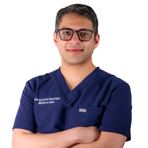
This page has been written and/or medically reviewed by Dr Sajjad Rajpar, Consultant Dermatologist and Medical Director of Midland Skin, Birmingham.
Dr Rajpar is on the GMC Specialist Register in Dermatology and has over 20 years’ experience in clinical dermatology. He provides clinical oversight to ensure the information on this page is accurate, balanced and consistent with current dermatology practice.
Dr Sajjad Rajpar
Date last updated: 26th Sep 2026
Book in with us
Expert, consultant led care in dermatology, skin surgery, eyelid surgery, lasers and aesthetics.
Disclaimer
The content on this website is provided for general information only. It is not, and must not be treated as, medical advice, a diagnosis, or a recommendation for any particular treatment. We make reasonable efforts to keep information accurate and up to date, but medicine and technology change, and content may not reflect the most current evidence, guidance, product information, or individual practice. You should not rely on this website to make decisions about your health or treatment. Always seek personalised advice from an appropriately qualified healthcare professional. Any examples, images, testimonials, and outcome descriptions are illustrative only. Results vary between individuals and no outcome can be guaranteed.

