
Melanoma treatment in Birmingham
Specialist melanoma screening, diagnosis, surgery and follow-up
We provide Consultant led melanoma screening, assessment of suspicious moles, diagnostic surgery, treatment for suitable melanomas and post-treatment follow-up. We see patients from Birmingham, Solihull, Coventry, Wolverhampton, Worcester and across the Midlands.


Quick Facts about melanoma

Best for
Changing mole or melanoma

Appointment
Mole check or surgical assessment

Treatment
Excision and follow-up
Symptoms
New or changing lesion
Provider
Consultant dermatologist

Price
See below
What is melanoma?
Melanoma is a type of skin cancer that develops from melanocytes, the cells that produce pigment within the skin.
It is less common than basal cell carcinoma and squamous cell carcinoma but is more likely to spread to other parts of the body if it is not identified and treated early.
A melanoma may develop within an existing mole or appear as a new mark on previously normal skin. Although many melanomas are brown or black, they can also be pink, red, pale or contain several different colours.
Melanoma in situ, also called stage 0 melanoma, is the earliest form. The melanoma cells remain within the top layer of the skin and have not become invasive or spread elsewhere. It can be cured by complete surgical removal, but it may develop into invasive melanoma if it is not treated.
T1a melanoma is an early invasive melanoma measuring less than 0.8 mm in Breslow thickness without ulceration.
Melanoma in situ and T1a melanoma can usually be treated successfully with surgery.
Who is more likely to develop melanoma?
People may have an increased risk if they:
- have fair skin that burns easily, freckles, light hair or light-coloured eyes
- have had severe or repeated sunburn, particularly during childhood or early adulthood
- have used sunbeds
- have a large number of moles or several unusual-looking moles
- have previously had melanoma or another form of skin cancer
- have a close family member who has had melanoma
- have a weakened immune system
- have a large congenital mole or a recognised inherited melanoma syndrome
Melanoma can affect people of every skin colour and should not be dismissed because someone does not have the usual risk factors. In darker skin, melanoma may be more likely to develop on the palms, soles or beneath a nail.
What does melanoma look like?
There is no single appearance that identifies every melanoma. The most important warning sign is usually a new or changing lesion.
The ABCDE guide can help identify changes that should be assessed:
- A — Asymmetry: one half of the lesion looks different from the other
- B — Border: the edge is irregular, blurred or notched
- C — Colour: the colour is uneven or contains different shades of brown, black, red, pink, blue or white
- D — Diameter: many melanomas are larger than 6 mm, although melanoma can be smaller
- E — Evolving: the lesion is growing or changing in its size, shape, colour, surface or symptoms
Other warning signs include:
- a mole that looks noticeably different from your other moles
- a new pigmented or pink lesion that continues to grow
- bleeding, crusting, itching, tenderness or ulceration
- pigment spreading into the surrounding skin
- a new dark streak beneath a nail that is not explained by an injury
- a firm, raised lesion that is growing over weeks or months
Some nodular melanomas do not follow the usual ABCDE pattern. A lesion that is elevated, firm and growing should also be assessed.
When should I see a dermatologist?
Arrange a specialist assessment if:
- you have developed a new or changing mole
- a mole has altered in size, shape or colour
- one lesion looks different from your other moles
- a lesion is growing quickly or becoming raised and firm
- a mole or pigmented lesion is bleeding, crusting, itching or becoming sore
- you have developed an unexplained dark streak beneath a nail
- your GP or another clinician is concerned about a lesion
- you have previously had melanoma and have noticed a new or changing lesion
- you have multiple or atypical moles and would like a Consultant Dermatologist skin examination
- a biopsy has already confirmed melanoma in situ or invasive melanoma and you require further assessment or surgery
Melanoma is often painless. You should not wait for a lesion to become uncomfortable before arranging an assessment.

ken weston

Melanoma screening and mole checks at Midland Skin
Midland Skin offers Consultant Dermatologist-led mole checks and skin cancer screening.
Depending on the appointment arranged, this may involve assessment of a particular lesion or a wider examination of your skin. Please tell the office before booking if you would like a full skin examination or have several lesions you would like assessed so that the correct appointment and sufficient time can be arranged.
Your dermatologist will review your relevant medical history, previous skin cancers, family history, sun exposure and any changes you have noticed. Suspicious or unusual lesions are examined using a dermatoscope, which provides a magnified view of structures beneath the skin surface.
Clinical photographs may be taken as part of your medical record and to help plan or monitor treatment.
A skin check assesses the lesions present at that appointment. It cannot prevent a new melanoma from developing or guarantee that a lesion will not change later. Continuing to examine your own skin remains important.
Why getting the diagnosis right matters
Melanoma can resemble an ordinary mole, an atypical mole, seborrhoeic keratosis, pigmented basal cell carcinoma or another harmless or cancerous skin lesion.
Conversely, many irregular-looking moles are not melanoma.
Dermoscopic assessment helps the dermatologist decide whether a lesion appears reassuring, should be monitored or requires diagnostic surgery. A photograph, online assessment or automated mole scan cannot reliably confirm or exclude melanoma on its own.
When melanoma is suspected, the lesion should not be treated with laser, freezing or another destructive treatment because tissue is required for microscopic examination.
How is melanoma diagnosed?
Your Consultant Dermatologist examines the lesion, the surrounding skin and, where appropriate, the remainder of your skin. Dermoscopy and clinical photography are usually included.
If melanoma is suspected, the usual next step is a diagnostic excision. This involves numbing the skin with local anaesthetic and removing the entire visible lesion with a narrow margin of surrounding skin. The specimen is sent to a specialist pathology laboratory for examination under a microscope.
Diagnostic excision is normally arranged as a separate procedure rather than being carried out during the initial assessment.
If melanoma is confirmed, the pathology report provides information that determines the next stage of care. This includes:
- whether the melanoma is in situ or invasive
- the type of melanoma
- the Breslow thickness if the melanoma is invasive
- whether ulceration is present
- whether the initial excision margins are clear
- other features that may affect staging or management
The diagnosis and treatment plan cannot be confirmed until the histology result is available.
If melanoma has already been diagnosed elsewhere
You can arrange an assessment at Midland Skin if your melanoma in situ or invasive melanoma was diagnosed through the NHS or by another private provider.
Please provide your histology report, relevant clinic letters, photographs and clear information identifying the original biopsy site before your appointment. Additional pathology information or formal review of the original specimen may sometimes be required.
The biopsy site can become difficult to identify after it has healed, so an accurate photograph or diagram showing its original position is particularly valuable.
Your melanoma treatment pathway
Consultant assessment or mole screening
A Consultant Dermatologist examines the lesion or your skin using dermoscopy, reviews your risk factors and takes clinical photographs where appropriate.
Diagnostic excision and histology
If melanoma is suspected, the lesion is removed under local anaesthetic and sent for specialist histological examination. This is normally arranged as a separate procedure.
Results, staging and MDT planning
The histology determines whether the lesion is melanoma in situ or invasive melanoma and provides the information needed to stage it. Confirmed melanoma is managed through an appropriate multidisciplinary pathway.
Definitive treatment
Midland Skin regularly treats melanoma in situ and can provide wide local excision for suitable T1a melanomas. T1b, more advanced or complex melanomas are linked with an appropriate specialist skin cancer MDT for further investigation and treatment.
Results and follow-up
Your results, wound care and follow-up plan are arranged according to the final histology and melanoma stage. Follow-up may be provided at Midland Skin or shared with the specialist MDT.
How is melanoma treated at Midland Skin?
Surgery is the principal treatment for melanoma in situ and early invasive melanoma. The exact pathway depends on the histology, melanoma stage, position of the tumour, required reconstruction and your general health.
Diagnostic excision
Diagnostic excision removes a suspicious lesion so that the diagnosis and, where relevant, the melanoma thickness can be established accurately.
Although the first excision may appear to have removed the entire lesion, further surgery is normally required if melanoma in situ or invasive melanoma is confirmed.
Treatment for melanoma in situ
Midland Skin regularly diagnoses and treats melanoma in situ.
Melanoma in situ is also called stage 0 melanoma. The melanoma cells remain confined to the top layer of the skin and have not become invasive.
The standard treatment is a wide local excision. This removes an additional margin of skin from around and beneath the original biopsy scar to ensure the abnormal cells have been removed completely.
For melanoma in situ, the recommended clinical margin is usually at least 0.5 cm around the histological biopsy scar. The procedure is generally carried out under local anaesthetic, and the removed tissue is sent for further histological examination.
Depending on the size and position of the wound, it may be closed directly with stitches or repaired using a skin flap or skin graft.
Once melanoma in situ has been completely removed, no further cancer treatment is usually required.
Wide local excision for T1a melanoma
A T1a melanoma is a thin invasive melanoma measuring less than 0.8 mm in Breslow thickness without ulceration.
Midland Skin can provide definitive surgical treatment for suitable T1a melanomas. Suitability depends on the confirmed pathology, position of the melanoma, ability to identify the biopsy scar, the repair required and individual medical factors.
Treatment usually involves a wide local excision. A further clinical margin of skin is removed from around and beneath the original biopsy scar to reduce the risk of melanoma cells remaining in the surrounding skin.
For a stage I melanoma, the recommended clinical margin is usually 1 cm around the histological biopsy scar. The procedure is generally carried out under local anaesthetic, and the removed tissue is sent for further histological examination.
Depending on the size and position of the wound, it may be closed directly with stitches or repaired using a skin flap or skin graft.
More advanced or complex melanoma
If histology identifies a T1b or more advanced melanoma, or if the melanoma requires more complex investigation or surgery, we arrange involvement of an appropriate specialist skin cancer multidisciplinary team.
Depending on the stage and individual findings, the specialist pathway may include consideration of:
- sentinel lymph node biopsy
- lymph node ultrasound or other imaging
- more extensive or specialist reconstructive surgery
- oncology assessment
- immunotherapy, targeted treatment or radiotherapy
These treatments are not required for every melanoma. The appropriate investigations and treatment are determined by the specialist MDT.
Midland Skin can liaise with the treating hospital or oncology team and provide agreed post-treatment dermatology surveillance. Where care is shared, the responsibilities for clinical examinations, scans and oncology treatment will be made clear.
Follow-up after melanoma treatment
Midland Skin provides post-treatment follow-up for patients treated at the clinic and can also follow patients whose melanoma treatment was completed through the NHS or another private provider.
Follow-up may include:
- examination of the melanoma scar
- examination of the relevant regional lymph nodes
- a wider skin examination for another melanoma or skin cancer
- dermoscopy of relevant moles
- advice about skin self-examination and sun protection
- liaison with the treating specialist MDT
The follow-up schedule depends on the melanoma stage, histology and individual risk factors.
Routine scans and blood tests are not normally required after completely treated melanoma in situ or T1a melanoma. Any imaging required for more advanced melanoma is arranged according to the specialist MDT follow-up plan.
Checking your skin and reducing further risk
Once you have had melanoma in situ or invasive melanoma, there is an increased risk of developing another melanoma or another form of skin cancer.
Check your skin approximately once a month. Look at the whole skin surface, including the scalp, back, soles, between the toes and beneath the nails. A partner, mirror or a set of baseline photographs can help with areas that are difficult to see.
Arrange an assessment if you notice a new lesion, a changing mole, a change around the previous melanoma scar or an unexplained lump.
You can reduce further ultraviolet damage by:
- avoiding sunbeds
- avoiding sunburn
- using clothing, a hat and shade as your main forms of protection
- spending time out of strong midday sunlight
- applying a broad-spectrum SPF 30 or higher sunscreen generously to exposed skin
- reapplying sunscreen regularly and after swimming or heavy sweating
Sunscreen provides additional protection but does not replace clothing and shade.
Why choose Midland Skin for melanoma care?
Midland Skin provides a consultant-led pathway from screening and diagnosis through to early melanoma surgery and follow-up.
- Mole checks and suspicious lesions are assessed by Consultant Dermatologists.
- Dermoscopy and clinical photography are available as part of assessment.
- Diagnostic excision and histological examination can be arranged.
- Melanoma in situ is regularly diagnosed and treated at Midland Skin.
- Suitable T1a melanomas can be treated with wide local excision.
- You meet the clinician accepting responsibility for your procedure before surgery.
- Established MDT links provide a pathway for more advanced or complex melanoma.
- Post-treatment dermatology follow-up can be provided in liaison with the treating MDT.
- Care is provided within a CQC-registered Birmingham clinic.
Melanoma assessment and treatment: FAQs
Which appointment should I book?
Please tell the office whether you are concerned about one particular lesion, would like a wider mole check or already have a confirmed melanoma in situ or invasive melanoma.
This allows the correct appointment type and sufficient time to be arranged. If you have a histology report or clinic letter, please provide it before booking.
Can melanoma be diagnosed from a photograph?
A photograph can help with initial triage but cannot reliably confirm or exclude melanoma.
A face-to-face examination with dermoscopy is required. If the lesion remains suspicious, diagnosis depends on removing tissue and examining it under a microscope.
Can a suspicious mole be removed at my first appointment?
Assessment and diagnostic surgery are normally arranged as separate appointments.
The clinician accepting responsibility for the procedure must assess the lesion, confirm the treatment site, consider the appropriate biopsy method and plan the wound repair and histology before surgery.
I already have a confirmed melanoma. Can Midland Skin treat it?
Yes. Midland Skin regularly treats melanoma in situ and can provide wide local excision for suitable T1a melanomas.
More advanced or complex melanomas are linked with an appropriate specialist MDT. Please send your histology report, clinic letters and photographs before the appointment.
Why do I need more surgery if the first excision had clear margins?
The first operation is a diagnostic excision performed with a narrow margin. If melanoma in situ or invasive melanoma is confirmed, a wide local excision removes an additional safety margin from around and beneath the biopsy scar.
This reduces the likelihood of melanoma cells remaining in the nearby skin and lowers the risk of the melanoma returning at the original site.
Will I need a sentinel lymph node biopsy or scans?
Sentinel lymph node biopsy and routine staging scans are not normally required for melanoma in situ or T1a melanoma.
They may be considered for thicker or higher-risk melanoma depending on the Breslow thickness, ulceration, other pathological features and clinical findings. These decisions are made through the specialist melanoma MDT.
Can Midland Skin provide follow-up if my treatment was carried out elsewhere?
Yes. We can assess patients who have completed melanoma treatment through the NHS or another private provider.
Where appropriate, Midland Skin can provide clinical skin, scar and lymph-node surveillance while liaising with the treating MDT. Any required imaging or oncology treatment remains coordinated through the specialist service.
Will melanoma surgery leave a scar?
Yes. Both diagnostic excision and wide local excision leave scars.
The wide local excision scar is usually longer than the original mole or biopsy scar because an additional treatment margin must be removed. Your Consultant will explain the likely wound, repair and recovery before you decide whether to proceed.
Fees
Specialist skin surgery assessment Show pricing
Book ConsultationThe consultation fee covers the Consultant’s assessment, examination, clinical photographs, clinical judgement and care plan. It remains payable whether surgery, referral, monitoring or no treatment is recommended. It is separate from, and is not deducted from, the fee for any subsequent procedure. If diagnostic or definitive surgery is recommended, the procedure and histology fees will be confirmed once the required treatment has been established. Fees for surgery include any immediate aftercare that is required, such as wound checks, dressing changes and stitch removal.
Initial dermatology assessment
Up to 25 minutes
Biopsy of skin
Skin cancer excision (per lesion)
Review consultation
Standard, up to 20 minutes
References used for this article
- National Institute for Health and Care Excellence. Melanoma: assessment and management (NG14). Updated July 2022.
- British Association of Dermatologists. Melanoma in situ patient information leaflet. Updated April 2026.
- British Association of Dermatologists. Melanoma stage 1A patient information leaflet. Updated October 2022.
- Garbe C, et al. European consensus-based interdisciplinary guideline for melanoma. Part 1: Diagnostics — Update 2024. Eur J Cancer. 2025;215:115152. doi:10.1016/j.ejca.2024.115152.
- Garbe C, et al. European consensus-based interdisciplinary guideline for melanoma. Part 2: Treatment — Update 2024. Eur J Cancer. 2025;215:115153. doi:10.1016/j.ejca.2024.115153.
About the Author
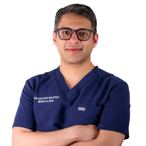
This page has been written and/or medically reviewed by Dr Sajjad Rajpar, Consultant Dermatologist and Medical Director of Midland Skin, Birmingham.
Dr Rajpar is on the GMC Specialist Register in Dermatology and has over 20 years’ experience in clinical dermatology. He provides clinical oversight to ensure the information on this page is accurate, balanced and consistent with current dermatology practice.
Dr Sajjad Rajpar
Date last updated: 22nd Sep 2026
Book in with us
Expert, consultant led care in dermatology, skin surgery, eyelid surgery, lasers and aesthetics.
Disclaimer
The content on this website is provided for general information only. It is not, and must not be treated as, medical advice, a diagnosis, or a recommendation for any particular treatment. We make reasonable efforts to keep information accurate and up to date, but medicine and technology change, and content may not reflect the most current evidence, guidance, product information, or individual practice. You should not rely on this website to make decisions about your health or treatment. Always seek personalised advice from an appropriately qualified healthcare professional. Any examples, images, testimonials, and outcome descriptions are illustrative only. Results vary between individuals and no outcome can be guaranteed.

